Category Archives: Academic

Deuteranopia – Red-Green Color Blindness

Deutan color vision deficiencies are by far the most common forms of color blindness. This subtype of red-green color blindness is found in about 6% of the male population, mostly in its mild form deuteranomaly.

Deuteranopia Color Spectrum
Normal and Deuteranopia Color Spectrum

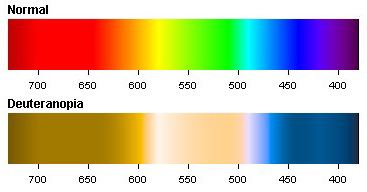
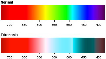
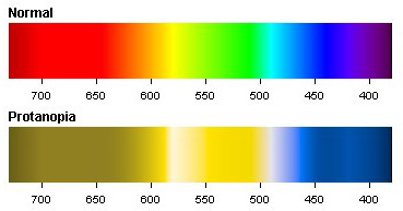
When you have a look at the color spectrum of a deuteranopic person you can see that a variety of colors look different than in a normal color spectrum. Whereas red and green are the main problem colors, there are also for example some gray, purple and a greenish blue-green which can’t be distinguished very well.

The well known term red-green color blindness is actually split into two different subtypes. On one side persons which either lack or have anomalous long wavelength sensitive cones (protan color vision deficiency), which are more responsible for the red part of vision. And on the other side deutan color vision deficiencies, which again are split into two different types:

  1. Dichromats: Deuteranopia (also called green-blind). In this case the medium wavelength sensitive cones (green) are missing at all. A deuteranope can only distinguish 2 to 3 different hues, whereas somebody with normal vision sees 7 different hues.
  2. Anomalous Trichromats: Deuteranomaly (green-weak). This can be everything between almost normal color vision and deuteranopia. The green sensitive cones are not missing in this case, but the peak of sensitivity is moved towards the red sensitive cones.

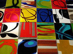
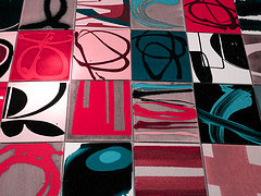
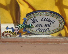
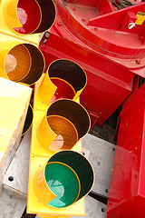
Below you can see a picture with normal colors on the left side and altered colors on the right side. The picture on the right side shows you how a person affected by deuteranopia would see the scenery (picture taken by Ottmar Liebert, some rights reserverd).
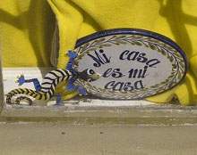
Normal Vision Deuteranopic Vision

In the midst of the last century there were different researches published concerning unilateral deuteranopia. Some persons were found which had trichromatic vision in one eye and dichromatic vision in the other. The eye with dichromatic vision had a color specturm related to a deuteranopia color spectrum. One case of such a one-eyed colorblind is described in the article The Spectral Luminosity Curves for a Dichromatic Eye and a Normal Eye in the Same Person.

The one-eyed color blindness is definitely not the common case, whereas deuteranopia and especially deuteranomaly are the most observed cases of all color vision deficiencies. In 75% of all occurrences of color blindness it is a defect caused by the green sensitive cones. The following list shows the approximative rates of deutan defects in our population:

  1. Deuteranomaly, Male Population: 5%
  2. Deuteranopia, Male Population: 1%
  3. Deuteranomaly, Female Population: 0.35%
  4. Deuteranopia, Female Population: 0.1%

These numbers don’t change much, because deutan color blindness as one form of red-green color blindness is a congenital disease. Red-green color blindness is a sex-linked trait and therefore encoded on the X chromosome. Because women have two X and can overcome the handicap of one, men have only one and are therefore more often affected. This circumstance can also be read in the numbers of the table above. More details about the concrete inheritance pattern can be found at The Biology behind Red-Green Color Blindness.

If you are colorblind there is a big chance that you are red-green colorblind, usually green-weak and male. And if you are suffering from deuteranomaly I just want to let you know, that you are nothing special…

Read more about Tritanopia and Protanopia—the other two types of color blindness.

The Way to Cure Color Blindness?

As for today there is no known treatment to cure color blindness but maybe in the near future there will be some possibilities to overcome color blindness and even enhance color vision overall.

Colorblind Mouse
Colorblind Mouse by Beige Alert

Researchers from the University of California and the Johns Hopkins Medical School in Baltimore showed in a series of experiments, how the color vision abilities of mice can be enhanced. Mice have naturally only dichromatic vision which can be compared to red-green color blindness. Dichromatic means, they have only two different types of color receptors in their eyes whereas humans normally have three different types which make up our color vision.

The scientists “pimped” the mice in their experiments with the missing genes for color perception which they took from humans. And those mice performed much better in the test setup as their dichromatic colleagues. This led to the assumption that the enhanced mice have a better (trichromatic) color vision.

Other researchers have also shown that they could cure color blindness in monkeys through injections of the missing color receptor genes.

What can we learn from this?

  • Color Blindness can be cured or at least it could be cured in about 6 cases of mice and monkeys so far.
  • We are on the way of the perfect human being and don’t even forget to exterminate color blindness on that way.
  • You won’t experience the cure of color blindness because this are only the first steps from a very long way.

If you are looking for a treatment of color blindness you still need to wait quite a while. And in case you can’t wait—hope dies last.

The Washington Post published an article about this research last week called Mice See New Hue With Added Gene. The original work was published in the science magazine: Emergence of Novel Color Vision in Mice Engineered to Express a Human Cone Photopigment.

Also read more about some newer scientific results on the cure of color-blind monkeys by gene therapy.

Terminology of Color Blindness

If you scratch under the surface of color blindness you sooner or later will come across many different terms related to color blindness which are not really self-explanatory. To get a better understanding of the terminology of color blindness I try to lift the curtain at least a bit.

General terms
Color blindness – a term which is misleading – is also known as color vision deficiency or daltonism. Color vision deficiency is not very well known but describes the phenomenon more precisely. Daltonism is named after the first scientist who wrote about color blindness. More details about these terms can be read in my recent article about Color Blindness is not ‘Color Blindness’.

Types of color blindness
There are four different types of color blindness which can be distinguished. This relates to the fact, that humans have three different color receptors in the eye (red, green and blue sensitive cones) and each of them can either be absent or working not properly. The fourth type describes the real color blindness.

  • Protan: The first type of color blindness relates to the red cones. Protanopia describes the fact that these cones are missing at all whereas protanomaly describes a displacement of them. Better terms would be red-blind as a synonym for protanopic and red-weak as a synonym for protanomalous.
  • Deutan: This term describes all green cone related conditions. If the green cones are missing it is called deuteranopia and a displacement is called deuteranomaly. Again some better terms to describe theses deficiencies in common speech would be green-blind (deuteranopic) and green-weak (deuteranomalous).
  • Tritan: Blue cone deficiencies are either called tritanopia if the blue sensitive cones are absent or tritanomaly if they are displaced. According to red and green cone deficiencies tritanopic is also called blue-blind and tritanomalous blue-weak.
  • Achromatopsia: This is the real color blindness. An other term for achromatopsia is rod monochromacy, because the cones are almost completely missing and since cones see colors whereas rods only see lightness this relates to complete color blindness and even a strong sensitiveness to bright light.

The very well known term red-green color blindness is an umbrella term including protan and deutan vision deficiencies. More information can be found in my earlier articles about protanopia and tritanopia.

Types of color vision
Color vision can be different in animals than in humans. Some animals have more types of cone cells and some have less. Even humans can not only have less but also more than three color receptors (see the article about tetrachromats).

  • Tetrachromatism: Four different color receptors. This is very unusual in humans but can be found in some animals.
  • Trichromatism: Three different color receptors related to red, green and blue. This is what we call normal vision.
  • Anomalous trichromatism: Three different color receptors whereas one of them is more or less good working. This relates to protanomaly, deuteranomaly and tritanomaly.
  • Dichromatism: Two different color receptors which describes the three different types of color blindness protanopia, deuteranopia and tritanopia.
  • Monochromatism: Either no color receptors at all or only one type of color receptors. This is also called rod monochromacy or achromatopsia.

These most commonly used terms in color blindness are not really common speech and can hardly be remembered. I like the terms red-blind, red-weak, green-blind, green-weak, blue-blind and blue-weak definitely the most. But unfortunately they are not very well known.

At least the term red-green color blindness is very accurate, describes the most common form of color vision deficiency and is even very well known.

Confusion Lines of the CIE 1931 Color Space

This is the second of three parts on the color blindness test based on the confusion lines of the CIE 1931 color space. In this part of the trilogy I would like to introduce you the confusion lines of the CIE 1931 color space, which was introduced in part one.

The CIE 1931 color space, which is two-dimensional, reflects only hue and saturation, which make up together chromaticity. The third dimension – lightness – is not shown in the diagram. But this doesn’t really matter, because many considerations of colors don’t need lightness. When looking at confusion lines we also don’t need the third dimension and therefore, they can be shown very nicely on the chromaticity diagram.

Let us have a look at some historical facts about confusion lines:

  • In 1855 J. C. Maxwell said: “Find two for a colorblind undistinguishable colors. Mark them on the CIE diagram and draw a line through them. This line will connect all colors which can’t be told apart by the colorblind person. You then can find more lines and all of those lines are either parallel or meet in a single point.”
  • A. König analyzed in 1892 the confusion lines and the so-called intersection point (also called co-punctal point) on three persons affected by color blindness.
  • In the year 1935 F. H. G. Pitt did some further research and found the confusion lines and corresponding intersection points for protanopic and deuteranopic persons.
  • D. Farnsworth (1955) and L. C. Thomson & W. D. Wright (1953) completed the work by adding the results for tritanopic persons.

Many studies followed and up to today these confusion lines are the main source while constructing tests on color blindness.

Confusion Lines - Protanopia
Confusion Lines – Protanopia

If you have a look at the diagram on the right side you can see the confusion lines associated to protanopic (red-blind) persons. The colors connected by one line can’t be distinguished by a protanope. If you would draw another line through the co-punctal point (intersection point), all colors on that line would look the same to a red-blind person too.

You can also see that there is a line going through a point called W. This is the so called white-point. Of course white can be told apart from red, even by a colorblind. But we have to take into account that the chromaticity diagram doesn’t include lightness. This means all colors along a line need the correct lightness adjustment to be undistinguishable by each other. Otherwise a colorblind can see a difference evenso it would be only a difference in brightness and not a different color perception.

Confusion Lines - Deuteranopia
Confusion Lines – Deuteranopia

The diagram of lines for deuteranopes (green-blind) looks quite the same as for protanopes. Both types of color blindness share a strong confusion on red and green colors, therefore the name red-green color blindness.

You can also see, that the lines are not exactly the same. Especially the intersection point is outside the range of the visible colors.

Confusion Lines - Tritanopia
Confusion Lines – Tritanopia

The last diagram looks totally different. The shown lines are connecting undistinguishable colors for tritanopes (blue-blind). Because the intersection point is at the blue end of the color spectrum, the color perception is completely different to the ones of red- or green-blind persons.

When you have a close look at all three diagrams you can also see, that the count of confusion lines differs. This is due to the following fact: Each line shows the smallest difference between distinguishable colors. This means not only the colors on one line, but all the colors between two lines are undistinguishable by persons affected by a certain type of color blindness.

In numbers:

  • People with normal vision can differentiate 150 wavelengths of colors.
  • Red-blind persons can see around 17 different wavelenghts.
  • Green-blind persons are able to distinguish around 27 wavelengths.
  • Blue-blind persons have an even more restricted visual field in the color spectrum.

By the way: Confusion lines are also called isochromatic lines, because they show lines of the same color (to the colorblind). A more accurate term would be pseudo-isochromatic lines, which is often used in academical papers.

In part three of this series, which will be the last part, we will have a look at the color blindness test based on these lines

Further readings:
Fundamental Studies Of Color Vision from 1860 To 1960
The Perception of Color

This is the second part about the color blindness test based on confusion lines of the CIE 1931 color space. The first part can be found at: CIE 1931 Color Space and the last part at: Color Blindness Test based on Confusion Lines of the CIE 1931 Color Space.

CIE 1931 Color Space

I would like to introduce a color blindness test based on the confusion lines of the CIE 1931 color space (also known as CIE XYZ color space). Because the topic is not the easiest one and needs some explanation I would like to split it into three parts. In this first of three parts I will introduce the CIE 1931 color space. In part two and three which will follow next week I will talk about the confusion lines of the CIE 1931 color space and last but not least the color blindness test based on the CIE 1931 color space confusion lines.

CIE 1931 Color Space

The acronym CIE stands for International Commission on Illumination which is the international authority on light, illumination, color, and color spaces. And 1931 is the year of birth of this specific color space.

Where it all began

Let us go back into history and have a look at where it all began. Based on the research on wavelengths and colors of Thomas Young at the turn of the 19th century, Hermann von Helmholtz developed around fifty years later a color theory. He stated, that the human eye has three different types of color receptors (cones) and that every color we perceive is a mixture of signals of those three types of cones, which roughly reflect the three different colors red, green and blue.

In the roaring twenties of the last century two scientists (W. David Wright, John Guild) took up this theory and independently made some experiments. To find out more about the three-color-theory a setup with four colored lights on two different sides was used.

  • On the left side a test color was projected by a light source.
  • On the right side the observer had three adjustable light sources (red, green and blue).

Now each observer (also called CIE standard observer) had the task to adjust the three lights accordingly, that the color on the right hand side was exactly the same as the test color on the other side. After many tests with a lot of test persons the results were mathematically analyzed. This produced three different curves of intensity for each light source to mix all colors of the color spectrum.

Funnily enough not every color could be matched and sometimes some red had to be added to the test color to get a correct match. This was also taken into account of the mathematical equations and resulted in a red curve including negative values. These three curves were standardized and are called the CIE RGB color matching functions r, g and b.

CIE RGB to CIE 1931

Because mathematicians don’t like negative numbers if they can change it, the commission changed it. Based on the CIE RGB functions and their corresponding values, new functions were calculated. Those new functions called x, y and z had to fulfill a list of conditions. Some of them were:

  • The new functions must be everywhere greater or equal to zero.
  • The y function describes only the luminosity.
  • The white point is, where x = y = z = 1/3.

All this put together produced the well known CIE XYZ color space which is also known as CIE 1931 color space. This color space aims to describe all visible colors to the human eye and can be shown as a three dimensional cube.

Projection

Because three dimensional objects can’t be illustrated very well a two dimensional representation had to be found. The Y parameter of the so-called tristimulus values X, Y and Z is a measure of the brightness. This helped to easily calculate the new chromaticity values x and y by the following rules:

  • x = X / (X + Y + Z)
  • y = Y / (X + Y + Z)

The corresponding chromaticity diagram is shown in the above picture. The outer curved line is called spectral locus and corresponds to the well known color spectrum, shown with corresponding wavelengths. The straight line on the lower part between blue and red is called purple line. This line relates to all colors which can only be mixed up by blue and red which are not part of the color spectrum.

In the next part I will talk about the confusion lines of the CIE 1931 color spectrum. And this leads us to the final part about the color blindness test based on those confusion lines.

Protanopia – Red-Green Color Blindness

Red-green color blindness is split into two different types: Whereas people affected by protan color blindness are less sensitive to red light, deuteranopia or deuteranomly (the second type of red-green color blindness) is related to sensitiveness on green light.

Gender Protanopia Protanomaly
Male 1.01% 1.08%
Female 0.02% 0.03%
Ratios by Gender

Protans have either defective long-wavelength cones (L-cones) or the L-cones are missing at all. If they are missing it is called protanopia or sometimes red-dichromacy. Affected persons are dichromats because they have only two working cone types, short- and medium-wavelength, compared to persons with normal vision with three different cone types.

If the L-cones are defective they appear in different intensities. This results in either a stronger or a weaker color blindness. If L-cones are not missing but defective it is called protanomaly. People suffering from this kind of color blindness are called anomalous trichromats.

Protanopia Color Spectrum

Protans have difficulties to distinguish between blue and green colors and also between red and green colors. When comparing the two spectrums you can see that there are different colors and shades of colors which are hard to distinguish for a protanopic person. So those persons are not only blind on red and green colors but a lot more. This means the well known term red-green color blindness is actually misleading and gives a wrong impression of protan color blindness (and also deutan color blindness).

Protanopia and protanomaly both are congenital color vision deficiencies. Their cause is an unequal recombination in the gene array which is passed on thereafter from parents to their children.

The genes encoding the L-cone photopigments are located on the X chromosome. This chromosome is also called the sex-chromosome, because women have two X’s compared to men with only one X combined with Y chromosome. If something is encoded on the X chromosome it is called sex linked. Sex linked traits are more often observed on men than women because a woman always has a second X chromosome which can compensate the deficiency. This unbalance between men and women can be seen in the table above showing the ratios of each kind of protan color blindness.

Traffic Light
Traffic Light by Jay P.
Some rights reserved.

There are a number of studies which show that color vision deficiencies are a serious risk factor in driving. Particularly protan color blindness reduces substantially the ability to see red lights, regardless of the severity of the defect. Tests showed that protans were very much over-represented in an accident causing group of drivers mostly involving either signal lights or brake lights. Some scientists estimate that being a protan has associated with it a level of risk of road accident that is equivalent to having a blood alcohol level of between 0.05 and 0.08 per cent. Because of that for example in Australia you can’t get hold of a commercial drivers licence since 1994 if you are suffering from protanopia or protanomaly.

Read more about Tritanopia and Deuteranopia—the other two types of color blindness.

Further reading:
Opsin Genes, Cone Photopigments, Color Vision, and Color Blindness
Protan Colour Vision Deficiency and Road Accidents
Wikipedia: Color Blindness

Related articles:
The Biology Behind Red-Green Color Blindness
Colorblind Population
At The Traffic Light

Tritanopic after Head Injury

Divya from India asks me the following question. She tells a short story about acquiring color blindness after a heavy head injury. Here are the lines Divya sent to me:

my boyfriend suffered an injury while playin cricket recently and the optician diagnosed it as tritanopia..heres what happened..he was aplying and was kinda nauseous and he puked in his helmet and fainted and fell backwards and got a hard blow on his head from the bat and a whack from the ground at the back of his head again and got knocked out/fainted..he was out for almost an hour and when he regained consciousness he was colourblind…the optician said its tritanopia..and he can only see luminous stuff as in highlighters and violet stuff and other stuff appears gray…what i want to know is…is this permanent or is it temporary…and if it is curable how can it be done…

I will try my best to answer the above questions and give some more insights about acquired color blindness. As I am not a professional in the field of color vision professional help should be frequented for detailed clarifications.

Unfortunately color blindness can be caused by severe head injuries. By far the most occurences of color blindness are congenital. In very rare cases people with perfect color vision abilities can be affected by an impaired vision or color vision deficiency after a brain trauma, a stroke or some other kind of severe head injury.

Such vision deficiencies are caused by a damage of the optical cortex. Several cases are described in scientific papers but there are no results concerning the cause of the loss of color vision. Some speculate, that the patient’s abnormality arises from partial destruction of the chromatic mechanism. The case described by Divya above about an acquired tritanopia may also occur, if the rod-cone mechanism is damaged.

Unfortunately as much acquired color blindness as congenital color blindness are not curable or at least there are no methods known to this day. As an acquired color blindness is without much doubt caused by some damage in the optical cortex most likely there will be no cure in sight in the near future.

Also because most often a severe damage causes the color blindness, there is evidence to suggest that this trait will be permanent and not only a temporarly impairment.

Further reading:
Traumatically acquired color vision defect

Related article:
Tritanopia – Blue-Yellow Color Blindness

Chromosomes Involved in Color Blindness

Human beings have 23 pairs of chromosomes. Out of these 23 pairs 22 are autosomal chromosomes which are equal in both sexes and encode body functions. Only one pair consists of two sex-chromosomes which are different for men and women. The 22 pairs of equal chromosomes are numbered from 1 through to 22. The sex-chromosomes are labeled with X and Y, whereas women carry the combination XX and men the combination XY. This all sums up in a total of 46 chromosomes which make the human genome.

Karyotype
Human Karyotype

Color blindness was actually the trigger to start mapping the human genome. It all began in 1911, when red-green color blindness was assigned to the X chromosome. This was based upon the observation that color blindness is passed from mothers to their sons. Thereby the women are usually not affected because of the normal copy, the second X chromosome. Men in contrary can not oversteer the defective chromosome, because they are carrying just one X chromosome.

The project to decipher the whole human genome is these days much more advanced. Scientists are working eagerly to encode the whole approximately 30’000 genes in the human genome.

If we have a closer look at the chromosomes which are involved into color blindness, we should distinguish between the different types of color blindness because they are encoded at different places in the genome.

  • Red-green color blindness
    This term combines four different types of color blindness. Protanomaly and protanopia are caused by defective or even missing L-cones (long-wavelengths). In opposite defective or missing M-cones (medium-wavelengths) are the source of deuteranomaly or deuteranopia. The genes encoding the L- and M-cone photopigments are located side by side on the X chromosome. Because of the genes are highly homologous and adjacent to one another, recombinations between them is common and can lead to anomalous pigments.
  • Blue cone monochromacy
    As this type of monochromacy is caused by a complete absence of L- and M-cones, blue cone monochromacy is encoded at the same place as red-green color blindness on the X chromosome.
  • Blue-yellow color blindness
    Tritanomaly and tritanopia which are commonly referred to as blue-yellow color blindness are caused by defective or missing S-cones (short-wavelength). These photopigments are encoded in genes which reside on chromosome 7, an autosomal chromosome. This is why blue-yellow color blindness occures at the same rate on both sexes.
  • Rod monochromacy
    The total loss of color vision is called rod monochromacy or complete achromatopsia. In this case the retina does not have any cone cells at all. It is known to be an autosomal recessive disease and can be provoked by different circumstances. Recent studies show that it can be encoded on chromosome 2 as well as on chromosome 8. Earlier studies assigned chromosome 14 to rod monochromacy but this could not be reconstructed.

The genes encoding L-, M- and S-cone photopigments are very well understood and determined whereas the source of rod monochromacy is a topic which still needs further research. Supposably different circumstances can cause rod monochromacy.

Type Chromosome
Deuteranomaly X Chromosome
Deuteranopia X Chromosome
Protanomaly X Chromosome
Protanopia X Chromosome
Tritanomaly Chromosome 7
Tritanopia Chromosome 7
Blue Cone Monochromacy X Chromosome
Rod Monochromacy Chromosome 2/8

The table on the left hand side shows on a glance the different types of color blindness and their related chromosomes. We have at least 4 different chromosomes out of the 23 pairs which can be the source of color vision deficiencies. Further studies of the human genome will show which chromosomes carry the encoding genes for rod monochromacy as this is still a subject under research and therefore this table will maybe undergo some adjustements in the near future.

Further readings:
Genetics Home Reference: Chromosomes
National Center for Biotechnology Information Map Viewer
Homozygosity mapping of achromatopsia to chromosome 2
A locus for autosomal achromatopsia on human chromosome 8

Related articles:
Tritanopia – Blue-Yellow Color Blindness
The Biology behind Red-Green Color Blindness
Daltonism – Named after John Dalton

If you did like this article subscribe to the feed or newsletter of Colblindor.

Tritanopia – Blue-Yellow Color Blindness

Actually the wording blue-yellow color blindness is misleading. People affected by tritan color blindness confuse blue with green and yellow with violet. So the term blue-green color blindness would be more accurate because the colors blue and yellow are usually not mixed up by tritanopes.

Tritan defects affect the short-wavelength cone (S-cone). There are two different types which can be observed:

  • Tritanopia: People affected by tritanopia are dichromats. This means the S-cones are completely missing and only long- and medium-wavelength cones are present.
  • Tritanomaly: This is an alleviated form of blue-yellow color blindness, where the S-cones are present but do have some kind of mutation.

Blue-yellow color blindness can be observed only very rarely. Different studies diverge a lot in the numbers but as a rule of thumb you could say one out of 10’000 persons is affected at most. In contrary to red-green color blindness tritan defects are autosomal and encoded on chromosome 7. This means tritanopia and tritanomaly are not sex-linked traits and therefore women and men are equally affected.

Tritanopia Color Spectrum
Normal and Tritanopia Color Spectrum

It can be observed that tritanopes usually have fewer problems in performing everyday tasks than do those with red-green dichromacy. Maybe this is because our society associates green with good/go and red with bad/stop, a pair of colors which accompanies us every day but a clear reason isn’t found yet by the researchers.

Tritan defects can not only be inherited but also acquired during one’s lifetime. In this case it even may be reversible and not permanent like an inherited color blindness. In the case of an acquired defect this is either evolving slowly for example simply through aging or coming instantly caused by a hard hit on your head.

  • Because the eye lens becomes less transparent with age, this can cause very light tritanomalous symptoms. Usually they are not serious enough for a positive diagnosis on color blindness.
  • Among alcoholics a higher incidence rate of tritanopia could be counted. Large quantities of alcohol resulted in poorer color discrimination in all spectra but with significantly more errors in the blue-yellow versus the red-green color range.
  • Mixtures of organic solvents even at low concentrations may also impair color vision. Errors were measured mainly in the blue-yellow color spectrum.
  • An injury through a hard hit to the front of back of your head may also cause blue-yellow color blindness. An example story can be found at Tritanopic after Head Injury.

The two photographs below give you some impression what tritanopes see. On the left side the actual photograph is shown as it is seen by people with normal color vision. On the right side you see the tritan counterpart where you can spot how blue-yellow color blindness influences the view of colors.

Art Plates Art Plates - Tritanopia

Photograph taken by Ottmar LiebertSome rights reserved

Read more about Deuteranopia and Protanopia—the other two types of color blindness.

Further readings:
Opsin Genes, Cone Photopigments, Color Vision, and Color Blindness
Does Occupational Exposure to Organic Solvents Affect Colour Vision?
Wikipedia: Color Blindness

Related articles:
The Biology behind Red-Green Color Blindness
Colorblind Population

Colorblind Population

If you are suffering from red-green color blindness, what are the chances that your neighbour or one of your classmates also suffers from it? What we like are numbers and one of the most often asked and searched for questions about color vision deficiencies is: How many people are affected by color blindness?

Before we digg right into some interesting numbers about red-green color blindness you should be aware of the following facts:

  • Tritanopia (blue-yellow color blindness) is rare. Some sources estimate that 0.008% are affected by this type of color vision deficiency.
  • Monochromacy (complete color blindness) is very rare. Different sources vary between 1 in 33’000 to 100’000 (0.001%).
  • Families are not a good source of numbers concerning color blindness, because vision deficiencies are inherited and therefore some families are affected much more than others.

Red-green color blindness is the most common color vision deficiency; therefore most of the researches found regard this type of colorblind population. The following figures are listed in the book Color Vision: From genes to perception and combine many different surveys spread over the last century.

Gender Anomaly Anopia
Protan Deutan Protan Deutan
Male 1.08% 4.63% 1.01% 1.27%
Female 0.03% 0.36% 0.02% 0.01%

Ratios of red-green color blindness types

Four different deficiencies make up the common wording red-green color blindness. People suffering from an anomaly are trichromats but do report problems in color perception in the green-yellow-red sector of the spectrum. The two different types show a less sensitiveness either to red light (Protan) or green light (Deutan). In opposite people suffering from an anopia are dichromats, completely lacking one type of retinal cones.

The figures above basically show the following important facts about red-green color blindness:

  • Roughly 8% of men and 0.5% of women are affected. Therefore chances that your neighbour or one of your classmates is colorblind are very high.
  • Deutanomaly is by far the most common color vision deficiency regarding red-green color blindness. The other three types are occurring at nearly the same ratio and do affect about one out of 100 persons each.
  • Men are approximately 100 times more often affected than women. This shows very nicely that red-green color blindness is a sex-linked trait as described in more detail at The Biology behind Red-Green Color Blindness.

A last note about the figures: If you always thought that you are someone special because of your color blindness, you might be wrong. Especially if you are male and suffering from Deuteranonmaly one could say you are one under many and you can meet fellow sufferers out on the street every day.

Further readings:
Opsin Genes, Cone Photopigments, Color Vision, and Color Blindness
The Perception of Color

Related articles:
The Biology behind Red-Green Color Blindness
Color Blindness Test by Dr Shinobu Ishihara
5 Misbeliefs about Color Blindness